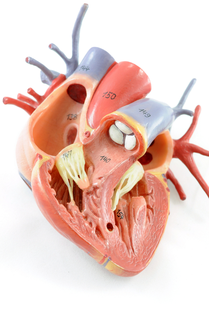
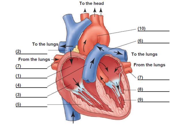
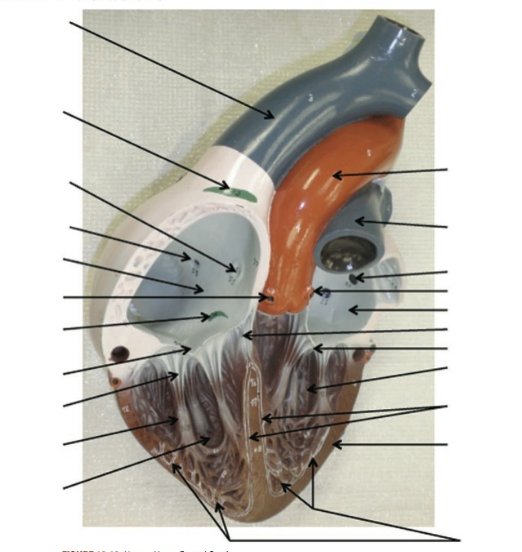
42 structure of the heart without labels
The Anatomy of the Heart - Quiz 1 - Free Anatomy Quiz The circulatory system - lower body image, with blank labels attached. The circulatory system - a PDF file of the upper and lower body for printing out to use off-line. Describe and explain the function of the circulatory system - The circulatory system consists of the heart, the blood vessels (veins, arteries, and capillaries), and the blood. Human Heart Diagram Without Labels - Labelling ... This fab labelling worksheet is a great way to help your kids with their learning of the basic human anatomy. It comes with a handy heart diagram without labels, so your students can label it themselves and familiarise themselves with the different parts of the human heart. This is a really useful way of complementing your teaching of the topic, giving pupils the chance to show what ...
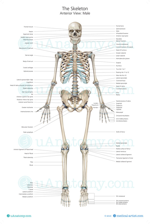
Human Heart Diagram Without Labels | Human heart diagram ... Human Heart Diagram Without Labels. Find this Pin and more on AnP by Susan Wells. This is Page 39 of a photographic atlas I created as a laboratory study resource for my BIOL 121 Anatomy and Physiology I students on the bones and bony landmarks of the axial skeleton. Credits: All photography, text, and labels by Rob Swatski, Assistant Professor ...
Structure of the heart without labels
Anatomy of a Human Heart - uofmhealth Anatomy of a Human Heart Author. Michigan Medicine. February 27, 2019 7:00 AM. Your heart does a lot of work to keep the body going. Learn about the organ's amazing power and the functions of its many parts. This story was updated on January 31, 2020. ... Human Heart Diagram Labeled - Science Trends The heart's atrioventricular valves are structures that join the atria and ventricles of the heart together. This group of valves is comprised of the tricuspid valve and the mitral valve. Beyond this, there is a structure referred to as the aortic valve which separates the left ventricle and the aorta. Heart Anatomy: Labeled Diagram, Structures, Blood Flow ... We will be using the 4 boxes to represent the chambers of the heart. Right vs Left Side of the Heart Now that we have converted the heart into a square with 4 different boxes or chambers, the heart can be divided into 2 sides. First, the right side is shown in blue and includes boxes/chambers 1 and 2.
Structure of the heart without labels. Heart Blood Flow | Simple Anatomy Diagram, Cardiac ... Step 2 involves the left atrium, the chamber of the heart that receives oxygenated blood from the lungs via the pulmonary veins. 3. Mitral Valve Step 3 involves the mitral valve. During diastole, when the heart is relaxed and filling with blood, the oxygenated blood from the left atrium will flow to the left ventricle. Heart Labeling Quiz: How Much You Know About Heart ... Here is a Heart labeling quiz for you. The human heart is a vital organ for every human. The more healthy your heart is, the longer the chances you have of surviving, so you better take care of it. Take the following quiz to know how much you know about your heart. Questions and Answers. 1. Human Heart Diagram Without Labels - Labelling Worksheet The human heart is a muscle made up of four chambers, these are: Two upper chambers - the left atrium and right atrium Two lower chambers - the left and right ventricles. It's also made up of four valves - these are known as the tricuspid, pulmonary, mitral and aortic valves. PDF Anatomy of Heart Labeled and Unlabeled Images (a) Anterior view of the external heart C' 2019 Pearson Education. Aort'c arch Ligamentum arteriosum Left pulmonary artery Left pulmonary ve ns Auricle of left atrium Circumflex artery Left coronary artery (in atrioventricular sulcus) Great cardiac vein Left ventricle Anterior interventricular artery (in anterior interventricular sulcus) Apex
PDF Anatomy of The Cardiovascular System COVERINGS OF THE HEART Structure of the Heart Coverings The heart has its own special covering, a loose-fitting inex-tensible sac called the pericardium. The pericardial sac, with the heart removed, can be seen in Figure 18-3. The peri-cardium consists of two parts: a fibrous portion and a serous portion (Figure 18-4). Label the heart - Science Learning Hub In this interactive, you can label parts of the human heart. Drag and drop the text labels onto the boxes next to the diagram. Selecting or hovering over a box will highlight each area in the diagram. Pulmonary vein Right atrium Semilunar valve Left ventricle Vena cava Right ventricle Pulmonary artery Aorta Left atrium Download Exercise Tweet Heart Diagram with Labels and Detailed Explanation - BYJUS Diagram of Heart. The human heart is the most crucial organ of the human body. It pumps blood from the heart to different parts of the body and back to the heart. The most common heart attack symptoms or warning signs are chest pain, breathlessness, nausea, sweating etc. The diagram of heart is beneficial for Class 10 and 12 and is frequently ... Heart Anatomy | Anatomy and Physiology - Lumen Learning The wall of the heart is composed of three layers of unequal thickness. From superficial to deep, these are the epicardium, the myocardium, and the endocardium. The outermost layer of the wall of the heart is also the innermost layer of the pericardium, the epicardium, or the visceral pericardium discussed earlier. Figure 6.
The Heart | Boundless Anatomy and Physiology The heart consists of four chambers separated into two sides. Each side contains an atria which receives blood into the heart and flows it into a ventricle, which pumps the blood out of the heart. The atria and ventricle on each side of the heart are linked together by valves that prevent backflow of blood. Human Heart - Diagram and Anatomy of the Heart The heart is a muscular organ about the size of a closed fist that functions as the body's circulatory pump. It takes in deoxygenated blood through the veins and delivers it to the lungs for oxygenation before pumping it into the various arteries (which provide oxygen and nutrients to body tissues by transporting the blood throughout the body). Structure of the Heart | Biology for Majors II In humans, the heart is about the size of a clenched fist, and it is divided into four chambers: two atria and two ventricles. There is one atrium and one ventricle on the right side and one atrium and one ventricle on the left side. The atria are the chambers that receive blood, and the ventricles are the chambers that pump blood. Structure of the Heart | SEER Training The human heart is a four-chambered muscular organ, shaped and sized roughly like a man's closed fist with two-thirds of the mass to the left of midline. The heart is enclosed in a pericardial sac that is lined with the parietal layers of a serous membrane. The visceral layer of the serous membrane forms the epicardium. Layers of the Heart Wall
Anatomy of the Heart - Medical Animation - YouTube This medical animation demonstrates the anatomy of the human heart, while explaining how the cardiovascular system functions. Explore more of our medical ani...
How the Heart Works - The Heart | NHLBI, NIH The Heart. The heart is an organ about the size of your fist that pumps blood through your body. It is made up of multiple layers of tissue. Your heart is at the center of your circulatory system. This system is a network of blood vessels, such as arteries, veins, and capillaries, that carries blood to and from all areas of your body.
Heart Anatomy: Labeled Diagram, Structures, Blood Flow ... We will be using the 4 boxes to represent the chambers of the heart. Right vs Left Side of the Heart Now that we have converted the heart into a square with 4 different boxes or chambers, the heart can be divided into 2 sides. First, the right side is shown in blue and includes boxes/chambers 1 and 2.
Human Heart Diagram Labeled - Science Trends The heart's atrioventricular valves are structures that join the atria and ventricles of the heart together. This group of valves is comprised of the tricuspid valve and the mitral valve. Beyond this, there is a structure referred to as the aortic valve which separates the left ventricle and the aorta.
Anatomy of a Human Heart - uofmhealth Anatomy of a Human Heart Author. Michigan Medicine. February 27, 2019 7:00 AM. Your heart does a lot of work to keep the body going. Learn about the organ's amazing power and the functions of its many parts. This story was updated on January 31, 2020. ...






Post a Comment for "42 structure of the heart without labels"