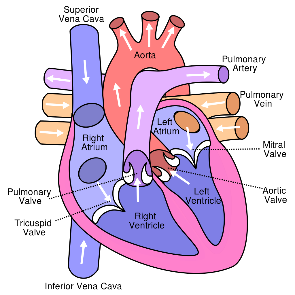
40 heart diagram with labels and blood flow
heart diagram blood flow heart diagram blood flow the Labeled Diagram Of The Heart And Blood Flow blood coming out of we have 9 Pics about the Labeled Diagram Of The Heart And Blood Flow blood coming out of like the Labeled Diagram Of The Heart And Blood Flow blood coming out of, Pin on College/ Nursing and also Pulmonary and Systemic Circulation - YouTube. Here you go: Practical notes - SP 2.3c Dissection of a Mammalian Heart 3. Use a glass rod to follow the path of blood flow : via the pulmonary vein, left atrium and through the bicuspid valve into the left ventricle; via the left ventricle through the semilunar valve and out of the aorta. 4. Note the muscular surface of the ventricle chambers which ensur es smooth blood flow. 5.
Circulatory System: Blood Flow Pathway Through the Heart Pathway of Blood Through the Heart. In this educational lesson, we learn about the blood flow order through the human heart in 14 easy steps, from the superior and inferior vena cava to the atria and ventricles. Come also learn with us the heart's anatomy, including where deoxygenated and oxygenated blood flow, in the superior vena cava, inferior vena cava, atrium, ventricle, aorta ...

Heart diagram with labels and blood flow
Expert Panel on Integrated Guidelines for Cardiovascular Health … In response, the former director of the National Heart, Lung, and Blood Institute (NHLBI), Dr Elizabeth Nabel, initiated development of cardiovascular health guidelines for pediatric care providers based on a formal evidence review of the science with an integrated format addressing all the major cardiovascular risk factors simultaneously. PDF BLOOD FLOW THROUGH THE HEART diagram BLOOD FLOW THROUGH THE HEART diagram BE ABLE TO EXPLAIN THE BLOOD FLOW THROUGH THE HEART: RIGHT SIDE (REMEMBER ANATOMICAL POSITION) LEFT SIDE DEOXYGENATED BLOOD FROM BODY TISSUE OXYGENATED BLOOD FROM LUNGS VIA PULMONARY VEINS SUPERIOR AND INFERIOR VENA CAVA LEFT ATRIUM RIGHT ATRIUM BICUSPID VALVE Sample 1: Heart and Lung Diagram - Accessible Image Sample Book The circulatory and respiratory systems work together to transport oxygen-rich blood through the body. A diagram shows a cross-section of a heart between two lungs. Red arrows show the path of oxygen-rich blood cells. Blue arrows show the path of oxygen-poor blood. Oxygen-rich blood cells travel to the heart from the lungs.
Heart diagram with labels and blood flow. Label the heart — Science Learning Hub Jun 16, 2017 · Labels. Description. Vena cava. Carries deoxygenated blood from the body to the heart. Semilunar valve. Flaps that prevent backflow of blood. Left atrium. Receives oxygenated blood from the lungs. Left ventricle. Region of the heart that pumps oxygenated blood to the body. Pulmonary artery. Carries deoxygenated blood to the lungs. Right ventricle 620 Human Heart Diagram Premium High Res Photos - Getty Images Browse 620 human heart diagram stock photos and images available, or search for heart illustration or pulmonary artery to find more great stock photos and pictures. Related searches: heart illustration. pulmonary artery. kidney diagram. Heart Anatomy: Labeled Diagram, Structures, Blood Flow ... - EZmed There are 4 chambers, labeled 1-4 on the diagram below. To help simplify things, we can convert the heart into a square. We will then divide that square into 4 different boxes which will represent the 4 chambers of the heart. The boxes are numbered to correlate with the labeled chambers on the cartoon diagram. How the Heart Works: Diagram, Anatomy, Blood Flow Illustrations of Blood Flow to the Heart Location and size of the heart The heart is located under the rib cage -- 2/3 of it is to the left of your breastbone (sternum) -- and between your lungs and above the diaphragm. The heart is about the size of a closed fist, weighs about 10.5 ounces, and is somewhat cone-shaped.
Diagram of Blood Flow Through the Heart - Bodytomy The heart is divided into two chambers, left and right, the right atrium and ventricle lie on the right side and the left atrium and ventricle on the left side. These two chambers are not directly connected to each other. Synchronization of the Two Chamber The right and left side or chambers of the heart work in tandem with each other. Diagram Of Body Organs Female Pics Pictures, Images and … Endometrial polyp or uterine polyp Endometrial polyp or uterine polyp. Sessile polyp and pedunculated polyp. The polyps consist of dense, fibrous tissue, blood vessels and endometrial epithelium. They are attached by a thin stalk or sessile. Vector diagram. diagram of body organs female pics stock illustrations PDF Arrows show the path of blood flow in the human heart. The blood enters ... Arrows show the path of blood flow in the human heart. The blood enters the heart from the body through the superior vena cava and the inferior vena cava. Then the blood enters the right atrium chamber of the heart. The blood then moves through the tricuspid valve (shown as two white flaps) into the right ventricle chamber of the heart. Heart anatomy: Structure, valves, coronary vessels - Kenhub The cusps are pushed open to allow blood flow in one direction, and then closed to seal the orifices and prevent the backflow of blood. Backward prolapse of the cusps is prevented by the chordae tendineae-also known as the heart strings-fibrous cords that connect the papillary muscles of the ventricular wall to the atrioventricular valves.. There are two sets of valves: atrioventricular ...
Label the heart - Science Learning Hub Interactive Label the heart Interactive Add to collection In this interactive, you can label parts of the human heart. Drag and drop the text labels onto the boxes next to the diagram. Selecting or hovering over a box will highlight each area in the diagram. Right ventricle Right atrium Left atrium Pulmonary artery Left ventricle Pulmonary vein Diagram of Human Heart and Blood Circulation in It Every heart diagram labeledwill clearly show these valves. These valves allow blood flow in one direction only. Different valves perform different functions. Tricuspid valve is located between the right ventricle of your heart and the right atrium, and allows the blood to move from the right atrium to the right ventricle. Heart Blood Flow Pictures, Images and Stock Photos Heart blood flow anatomical diagram with atrium and ventricle system. Vector illustration labeled medical poster. Blood circulation path scheme with arrows. Red Blood Cells Inside A Blood Vessel Red blodd cells inside a blood vessel. Horizontal composition with selective focus. Human bloodstream The human circulatory system Label Heart Anatomy Diagram Printout - EnchantedLearning.com The blood is then pumped to the left ventricle, then the blood is pumped through the aorta and to the rest of the body. This cycle is then repeated. Every day, the heart pumps about 2,000 gallons (7,600 liters) of blood, beating about 100,000 times. Label the heart anatomy diagram below using the heart glossary. Note: On the diagram, the right ...
The 8 Best Supplements To Boost Blood Flow Naturally - UMZU Dec 04, 2020 · Why Take A Nitric Oxide Boosting Supplement:. Increased Blood Flow & Circulation- This results in less cold extremities and improved nutrient/oxygen delivery.; Decreased Blood Pressure - Due to the expansion of the arterial walls during vasodilation, more blood is able to flow through the arteries at a lower blood pressure.; Improved performance - …
Blood Flow Through the Heart - Registered Nurse RN 1. The un-oxygenated blood (this is blood that has been "used up" by your body and needs to be resupplied with oxygen) enters the heart through the SUPERIOR AND INFERIOR VENA CAVA. 2. Blood enters into the RIGHT ATRIUM 3. Then it is squeezed through the TRICUSPID VALVE 4. Blood then enters into the RIGHT VENTRICLE 5.
Heart Diagram with Labels and Detailed Explanation - BYJUS The diagram of heart is beneficial for Class 10 and 12 and is frequently asked in the examinations. A detailed explanation of the heart along with a well-labelled diagram is given for reference. Well-Labelled Diagram of Heart The heart is made up of four chambers: The upper two chambers of the heart are called auricles.
Nervous System Worksheet Answers - WikiEducator Jan 14, 2008 · the activity of the heart and smooth muscle. A. Autonomic nervous system: 4. The part of the autonomic nervous system that increases heart and respiratory rates, increases blood flow to the skeletal muscles and dilates the pupils of the eye. E. Sympathetic nervous system: 5. The part of the autonomic nervous system that increases gut activity
Heart Blood Flow | Simple Anatomy Diagram, Cardiac Circulation Pathway ... Step 1 and 6 involve a blood vessel, which makes sense as this is how blood enters and exits that side of the heart. Steps 2-5 involve a chamber, valve, chamber, and valve. So if you remember this general pattern, it will help you recall the order in which blood flows through each side of the heart. Right Side of the Heart SVC/IVC Right Atrium
Blood Throught the Heart - Austin Community College District Blood Throught the Heart Blood Flow Through the Heart Beginning with the superior and inferior vena cavae and the coronary sinus, the flowchart below summarizes the flow of blood through the heart, including all arteries, veins, and valves that are passed along the way. 1. Superior and inferior vena cavae and the coronary sinus 2. Rt. atrium 3.
Circulatory System Diagram - Cardiovascular System and Blood ... SmartDraw has a number of templates included for circulatory system diagrams, cardiovascular system diagrams, blood circulation diagrams, and more. You don't really have to "draw" them as much as find them and modify them as needed. You can add labels or titles and change the size of symbols as necessary.
Pin on All Things Homeschool - Pinterest Shows a picture of a heart with a description of how blood flows through the heart, focusing on the chambers, vessels, and valves. Students are asked to label the heart and trace the flow of blood. Questions at the end of the activity reinforce important concepts about the heart and circulatory system.
Heart Information Center: Heart Anatomy - Texas Heart Institute The left ventricle's chamber walls are only about a half-inch thick, but they have enough force to push blood through the aortic valve and into your body. The Heart Valves. Four valves regulate blood flow through your heart: The tricuspid valve regulates blood flow between the right atrium and right ventricle.

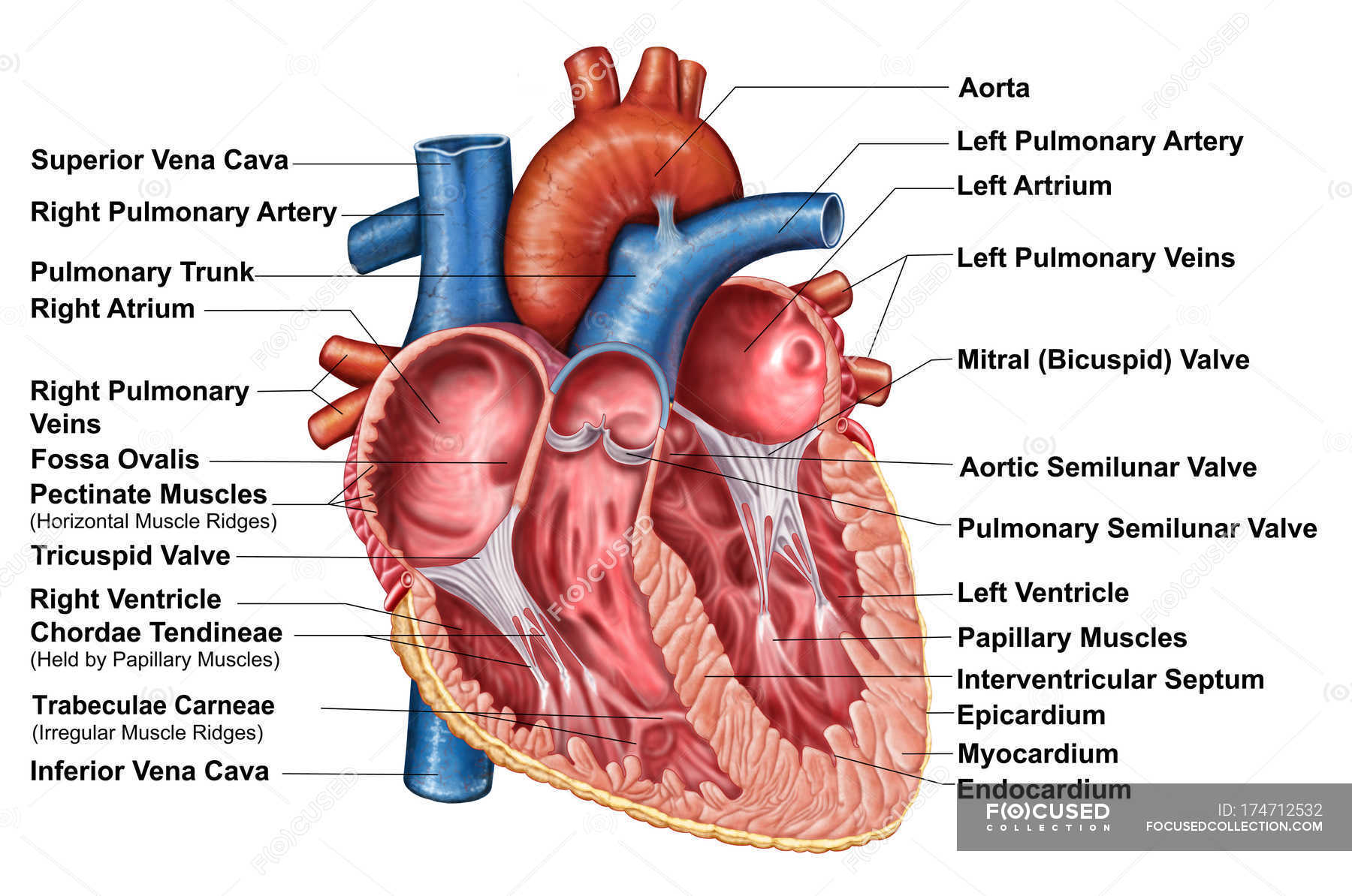
Anatomy of heart interior with labels — semilunar valve, pulmonary veins - Stock Photo | #174712532
The Heart Year 6 | KS2 Science | Twinkl (teacher made) Follow your heart and download this Animals Including Humans science lesson pack. This engaging resource enables children to learn about the three parts of the circulatory system and most specifically, the role of the heart. In this lesson, children recap on the names of organs that they have learnt in previous years and then they go on to learn about how the heart, blood …
Human Heart (Anatomy): Diagram, Function, Chambers, Location in ... - WebMD This lets blood flow better and can abort a heart attack or relieve angina (chest pain). Thrombolysis : “Clot-busting” drugs injected into the veins can …
YR 8 Topic 2 Circulatory System - Pinterest GREEN | Heart diagram, Human heart diagram, Circulatory system for kids From mrgscience.com YR 8 Topic 2 Circulatory System In this unit you will learn about how blood circulates through the body and how our bodies are supplied with energy. All cells in the body need to have oxygen and nutrients, and they need their... D Dawn Lovell Wilson
Human Heart - Anatomy, Functions and Facts about Heart Myocardial infarction is a serious medical condition where the blood flow to the heart is reduced or entirely stopped. This causes oxygen deprivation in the heart muscles, and prolonged deprivation can cause tissues to die. ... Label the Heart Diagram below: Practice your understanding of the heart structure. Drag and drop the correct labels to ...
A Diagram of the Heart and Its Functioning Explained in Detail The heart blood flow diagram (flowchart) given below will help you to understand the pathway of blood through the heart.Initial five points denotes impure or deoxygenated blood and the last five points denotes pure or oxygenated blood. 1.Different Parts of the Body ↓ 2.Major Veins ↓ 3.Right Atrium ↓ 4.Right Ventricle ↓ 5.Pulmonary Artery ↓ 6.Lungs
Human Heart Diagram Labeled | Science Trends Let's examine the anatomy of the heart along with some diagrams that show how the heart operates. Anatomy Of The Heart The human heart usually weighs somewhere between 10 to 12 ounces in men and between 8 to 10 ounces in women, and in terms of size is roughly the size of the fist.
PDF Heart Diagram Answer Key - University of Washington A: om the body: t lung VEINS: trium VENTRICLE: o the lung TRIUM VE A: o the body: o the lungs APEX VENTRICLE: o the body all VE TRIUM VEINS: trium e e A: om the body


.png)


Post a Comment for "40 heart diagram with labels and blood flow"