44 microscope images with labels
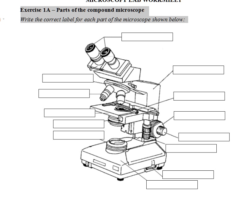
Binocular Microscope Anatomy - Parts and Functions with a Labeled ... Now, I will discuss the details anatomy of the light compound microscope with the labeled diagram. Why it is called binocular: because it has two ocular lenses or an eyepiece on the head that attaches to the objective lens, this ocular lens magnifies the image produced by the objective lens. Binocular microscope parts and functions Microscope Labeled Pictures, Images and Stock Photos Browse 49 microscope labeled stock photos and images available, or start a new search to explore more stock photos and images. Newest results Fluorescent Imaging immunofluorescence of cancer cells growing... Microscope diagram vector illustration. Labeled zoom instrument... Microscope diagram vector illustration.
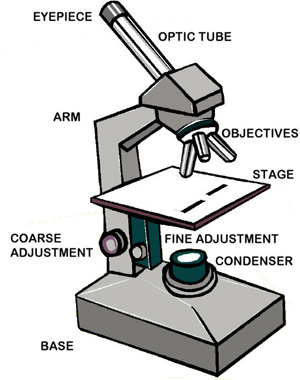
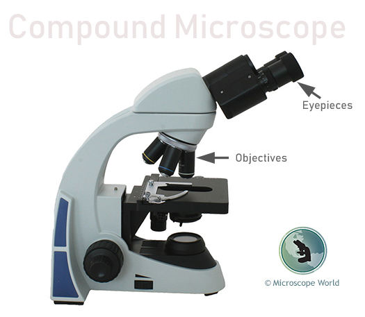
Compound Microscope Parts, Functions, and Labeled Diagram Compound Microscope Definitions for Labels. Eyepiece (ocular lens) with or without Pointer: The part that is looked through at the top of the compound microscope. Eyepieces typically have a magnification between 5x & 30x. Monocular or Binocular Head: Structural support that holds & connects the eyepieces to the objective lenses.

Microscope images with labels
18,701 Microscope drawing Images, Stock Photos & Vectors - Shutterstock Microscope drawing royalty-free images 18,701 microscope drawing stock photos, vectors, and illustrations are available royalty-free. See microscope drawing stock video clips Image type Orientation Color People Artists Sort by Popular Science Abstract Designs and Shapes College and University Art Styles Printing, Typography, and Calligraphy Index of Dr.Jastrow's electron microscopic atlas Scanning electron microscope: Scanning electron microscopy: E Material used in the course of Microscopic Anatomy at the University of Mainz, Germany which is provided by the WAI: digital electron microscopic images of c u r r e n t t e r m (in German) F Vocabulary of microscopic anatomy: Alphabetical index: Short information: Knowledge of cellular ultrastructure is an … Amazon.com: Magnifying Glasses 8X 15X 23X Magnifier LED … About this item . Double eye magnifying glasses magnifier loupe, with 2pcs adjustable LEDs to help it work in low-light conditions ; Left right double eye patches magnifierloupe with adjustable LED to help work in low-light conditions.Set of 2 magnifying glasses mounted on a one-size-fits-all eyeglass frame for easy hands-free operation.
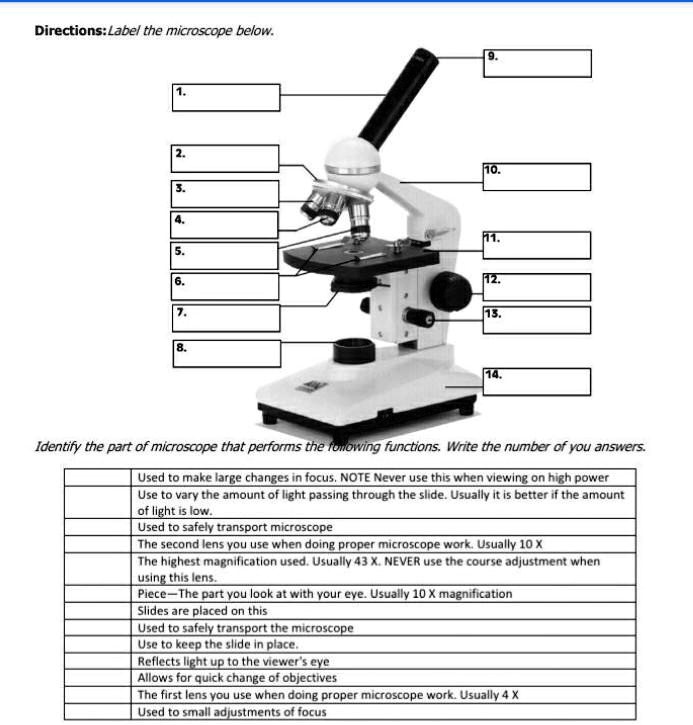
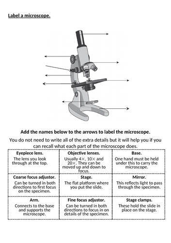
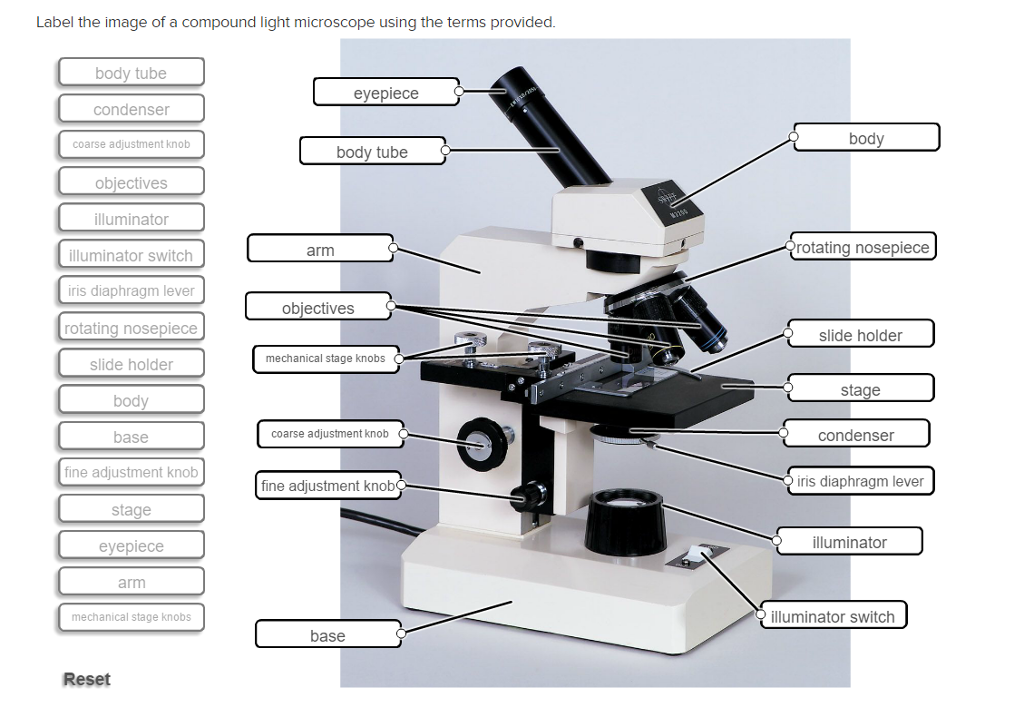
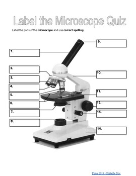
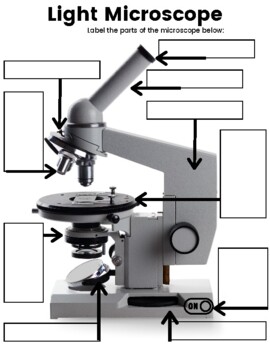
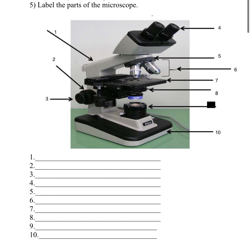
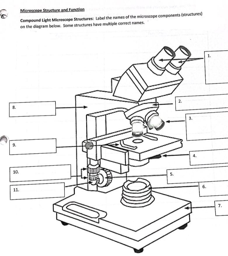
Microscope images with labels. Microscope Labeling Game - PurposeGames.com About this Quiz. This is an online quiz called Microscope Labeling Game. There is a printable worksheet available for download here so you can take the quiz with pen and paper. This quiz has tags. Click on the tags below to find other quizzes on the same subject. Science. Compound Microscope - Diagram (Parts labelled), Principle and Uses See: Labeled Diagram showing differences between compound and simple microscope parts Structural Components The three structural components include 1. Head This is the upper part of the microscope that houses the optical parts 2. Arm This part connects the head with the base and provides stability to the microscope. Parts of the Microscope with Labeling (also Free Printouts) Microscopes are specially created to magnify the image of the subject being studied. This exercise is created to be used in homes and schools. the microscope layout, including the blank and answered versions are available as pdf downloads. Click to Download : Label the Parts of the Microscope (A4) PDF print version. Mitosis Images Labeled | Virtual Anatomy Lab VAL - ncccval Endocrine Rabbit Dissection Unlabeled. Cardiovascular. Cardiovascular Histology Labeled. Cardiovascular Histology Unlabeled. Cardiovascular Models Labeled. Cardiovascular Models Unlabeled. Cardiovascular Sheep Heart Dissect-L. Cardiovascular Sheep Heart Disect-U. Cardiovascular Cat Dissection Labeled.
ZEISS Axioscope 5 Smart Laboratory Microscope Focus. Snap. Done. Forget about the 15 steps and clicks to document samples with multiple fluorescent labels. With Smart Microscopy, this is a thing of the past. Axioscope 5 with Axiocam 202 mono and Colibri 3 LED illumination take this workload from you. You keep your hands at the microscope stand. Relaxed. Labeling the Parts of the Microscope | Microscope World Resources Labeling the Parts of the Microscope This activity has been designed for use in homes and schools. Each microscope layout (both blank and the version with answers) are available as PDF downloads. You can view a more in-depth review of each part of the microscope here. Download the Label the Parts of the Microscope PDF printable version here. Microscope Labeling - The Biology Corner The google slides shown below have the same microscope image with the labels for students to copy. I often spend the first day walking students through the steps and having them look at a single slide as we do the steps. Students are often very enthusiastic about using microscopes and will try to start with the high power objective. Compound Microscope Parts - Labeled Diagram and their Functions The eyepiece (or ocular lens) is the lens part at the top of a microscope that the viewer looks through. The standard eyepiece has a magnification of 10x. You may exchange with an optional eyepiece ranging from 5x - 30x. [In this figure] The structure inside an eyepiece. The current design of the eyepiece is no longer a single convex lens.
Microscope Types (with labeled diagrams) and Functions The working principle of a simple microscope is that when a lens is held close to the eye, a virtual, magnified and erect image of a specimen is formed at the least possible distance from which a human eye can discern objects clearly. Simple microscope labeled diagram Simple microscope functions It is used in industrial applications like: Amazon.com : LCD Digital Microscope,ANNLOV 4.3 inch … Feb 05, 2020 · This item LCD Digital Microscope,ANNLOV 4.3 inch Handheld USB Microscope 50X-1000X Magnification Coin Microscope Video Camera with 8 Adjustable LED Lights for Adults PCB Soldering Kids Outside Use ANNLOV 7" LCD Digital Microscope with 32GB TF Card 1200X Maginfication 1080P Coin Microscope with Wired Remote,12MP Ultra-Precise Focusing Video ... Parts of Microscope Anatomy, Function, Names & Labeled Diagram Microscope parts labeled diagram gives us all the information about its parts and their position in the microscope. Microscope Parts Labeled Diagram The principle of the Microscope gives you an exact reason to use it. It works on the 3 principles. Magnification Resolving Power Numerical Aperture. Parts of Microscope Head Base Arm Eyepiece Lens PDF Label parts of the Microscope Label parts of the Microscope: . Created Date: 20150715115425Z
Intelligent microscope uses AI to capture rare biological events 20.09.2022 · The researchers used Micro-Manager software to capture images from the microscope and a neural network trained on labelled data to analyse them. For each image, the network output acts as a decision-making parameter to toggle between slow and fast imaging. Event recognition . To demonstrate their new technique, Manley and colleagues integrated EDA …
Electron Microscopy Images - Dartmouth We have a library of images recorded using our scanning and transmission electron microscopes. Some are shown below and others elsewhere. These images are in the public domain. If you have questions about the images or want some specific images contact Max Guinel . Hibiscus Flower (August 2021) Morphy Amorphophallus titanum anther cross section.
Label the microscope — Science Learning Hub All microscopes share features in common. In this interactive, you can label the different parts of a microscope. Use this with the Microscope parts activity to help students identify and label the main parts of a microscope and then describe their functions. Drag and drop the text labels onto the microscope diagram.
300+ Free Microscope & Laboratory Images - Pixabay 399 Free images of Microscope Related Images: laboratory science bacteria research scientist lab biology chemistry medical Find your perfect microscope image. Free pictures to download and use in your next project.
What is Electron Microscopy? - UMASS Medical School Because of its great depth of focus, a scanning electron microscope is the EM analog of a stereo light microscope. It provides detailed images of the surfaces of cells and whole organisms that are not possible by TEM. It can also be used for particle counting and size determination, and for process control. It is termed a scanning electron microscope because the image is formed by …
Parts of Stereo Microscope (Dissecting microscope) – labeled … Compared to a compound microscope where the objectives attached to the nosepiece can be seen and identified individually (based on color bands and their respective labels), the objectives of a dissecting microscope are located in a cylindrical cone and, therefore, are not directly seen. For the stereo microscope that comes with multiple objective lens sets (fixed power style), the …
Microscope Objective Lens | Products | Leica Microsystems The objective lens is a critical part of the microscope optics. The microscope objective is positioned near the sample, specimen, or object being observed. It has a very important role in imaging, as it forms the first magnified image of the sample. The numerical aperture (NA) of the objective indicates its ability to gather light and largely determines the microscope’s resolution, the ...
Simple Microscope - Diagram (Parts labelled), Principle, Formula and Uses A simple microscope consists of Optical parts Mechanical parts Labeled Diagram of simple microscope parts Optical parts The optical parts of a simple microscope include Lens Mirror Eyepiece Lens A simple microscope uses biconvex lens to magnify the image of a specimen under focus.
Microscope Parts and Functions First, the purpose of a microscope is to magnify a small object or to magnify the fine details of a larger object in order to examine minute specimens that cannot be seen by the naked eye. Here are the important compound microscope parts... Eyepiece: The lens the viewer looks through to see the specimen.
LAS X Industry Microscope software for Industry | Products Create sharp 2D images from several partially in-focus images. In connection with the 3D Surface Viewer, creation of 3D images is also possible. LAS X Stitching: Create 2D images from multiple tiled images captured automatically. Obtain a spiral scan to capture only the region which interests you most. Single images can be retrieved and ...
Explanation and Labelled Images - New York Microscope Company The samples are labeled with fluorophore where they absorb the high-intensity light from the source and emit a lower energy light of longer wavelength. The resulting fluorescent light is then separated from the surrounding radiation with filters, allowing the observer to see only the fluorescing material.
Electron microscope - Wikipedia An electron microscope is a microscope that uses a beam of accelerated electrons as a source of illumination. As the wavelength of an electron can be up to 100,000 times shorter than that of visible light photons, electron microscopes have a higher resolving power than light microscopes and can reveal the structure of smaller objects.. Electron microscopes use shaped magnetic …
Amazon.com: Magnifying Glasses 8X 15X 23X Magnifier LED … About this item . Double eye magnifying glasses magnifier loupe, with 2pcs adjustable LEDs to help it work in low-light conditions ; Left right double eye patches magnifierloupe with adjustable LED to help work in low-light conditions.Set of 2 magnifying glasses mounted on a one-size-fits-all eyeglass frame for easy hands-free operation.
Index of Dr.Jastrow's electron microscopic atlas Scanning electron microscope: Scanning electron microscopy: E Material used in the course of Microscopic Anatomy at the University of Mainz, Germany which is provided by the WAI: digital electron microscopic images of c u r r e n t t e r m (in German) F Vocabulary of microscopic anatomy: Alphabetical index: Short information: Knowledge of cellular ultrastructure is an …
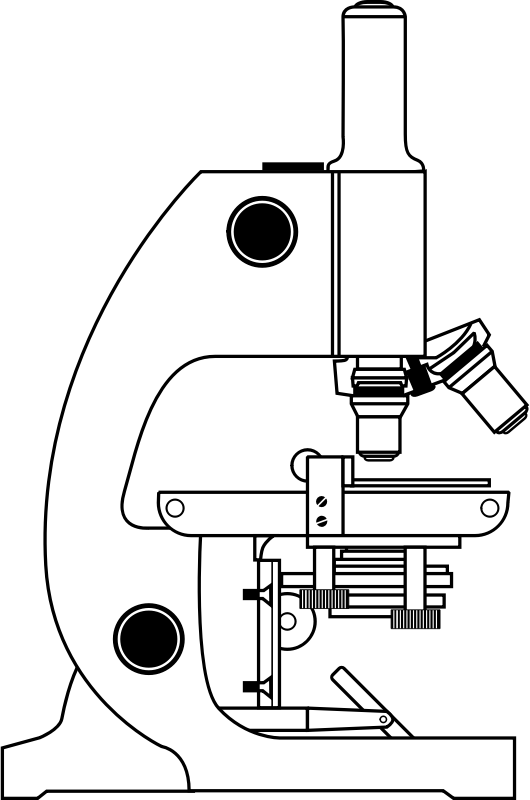
18,701 Microscope drawing Images, Stock Photos & Vectors - Shutterstock Microscope drawing royalty-free images 18,701 microscope drawing stock photos, vectors, and illustrations are available royalty-free. See microscope drawing stock video clips Image type Orientation Color People Artists Sort by Popular Science Abstract Designs and Shapes College and University Art Styles Printing, Typography, and Calligraphy













Post a Comment for "44 microscope images with labels"